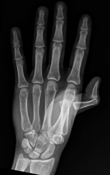
History/Physical Exam
The joint should be assessed for deformity, skin integrity, edema, and ecchymosis. Palpation may assist with determining whether the dislocation is dorsal or ventral. Dorsal dislocations have a palpable dorsal position of the proximal phalanx compared to the metacarpal, hyperextension of the MCP joint, and slight flexion of the IP joints. Overlapping of the digits while the patient makes a fist suggests a rotational component to the dislocation. A neurovascular exam, including sensation, capillary refill, and range of motion should be performed distal to the joint.
Associated ligamentous injury should be assessed after reduction, which demonstrates laxity with abduction or adduction while the joint is in flexion.