Patellar Tendon Rupture
Patellar Tendon Scanning
How to scan:
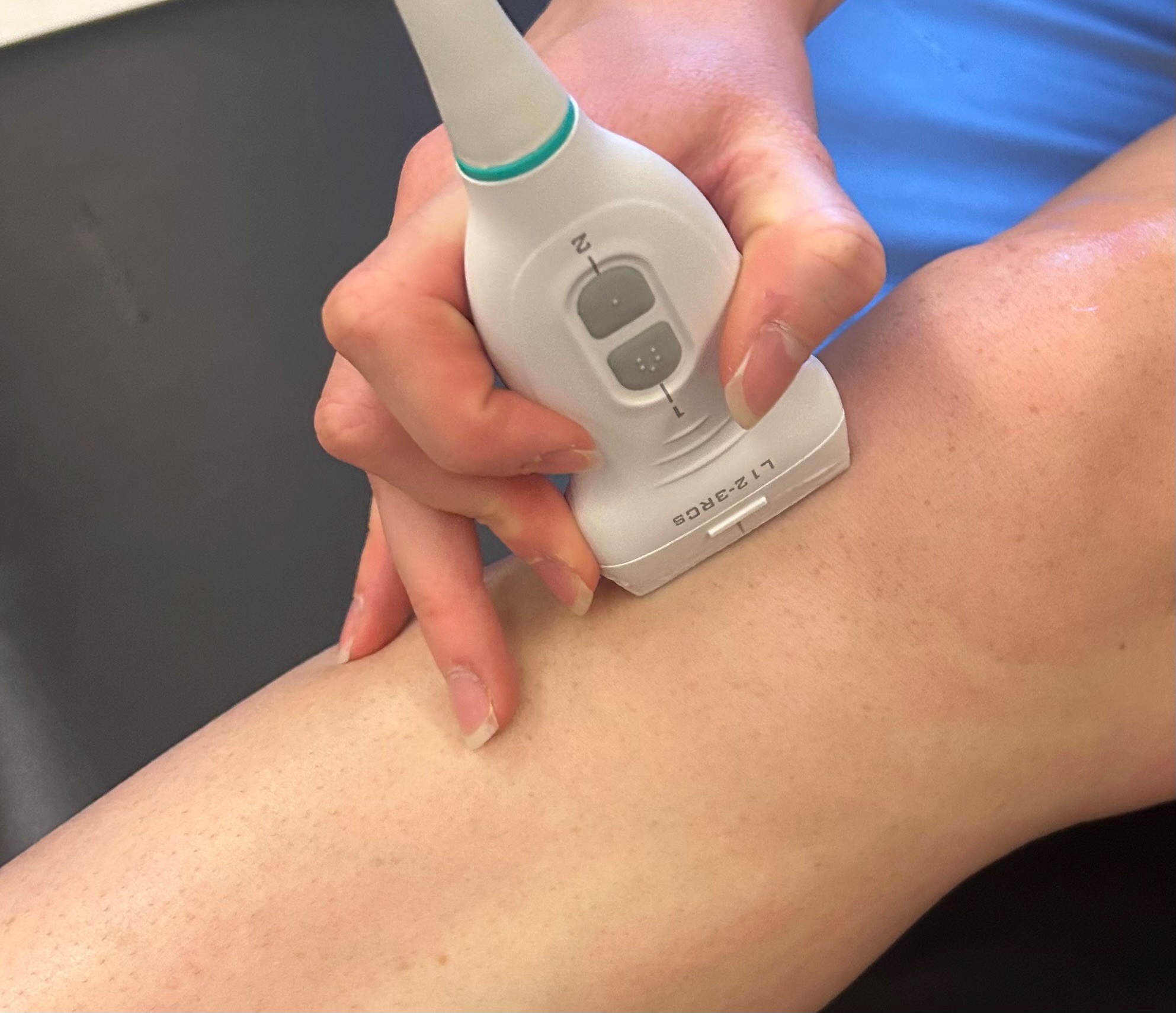
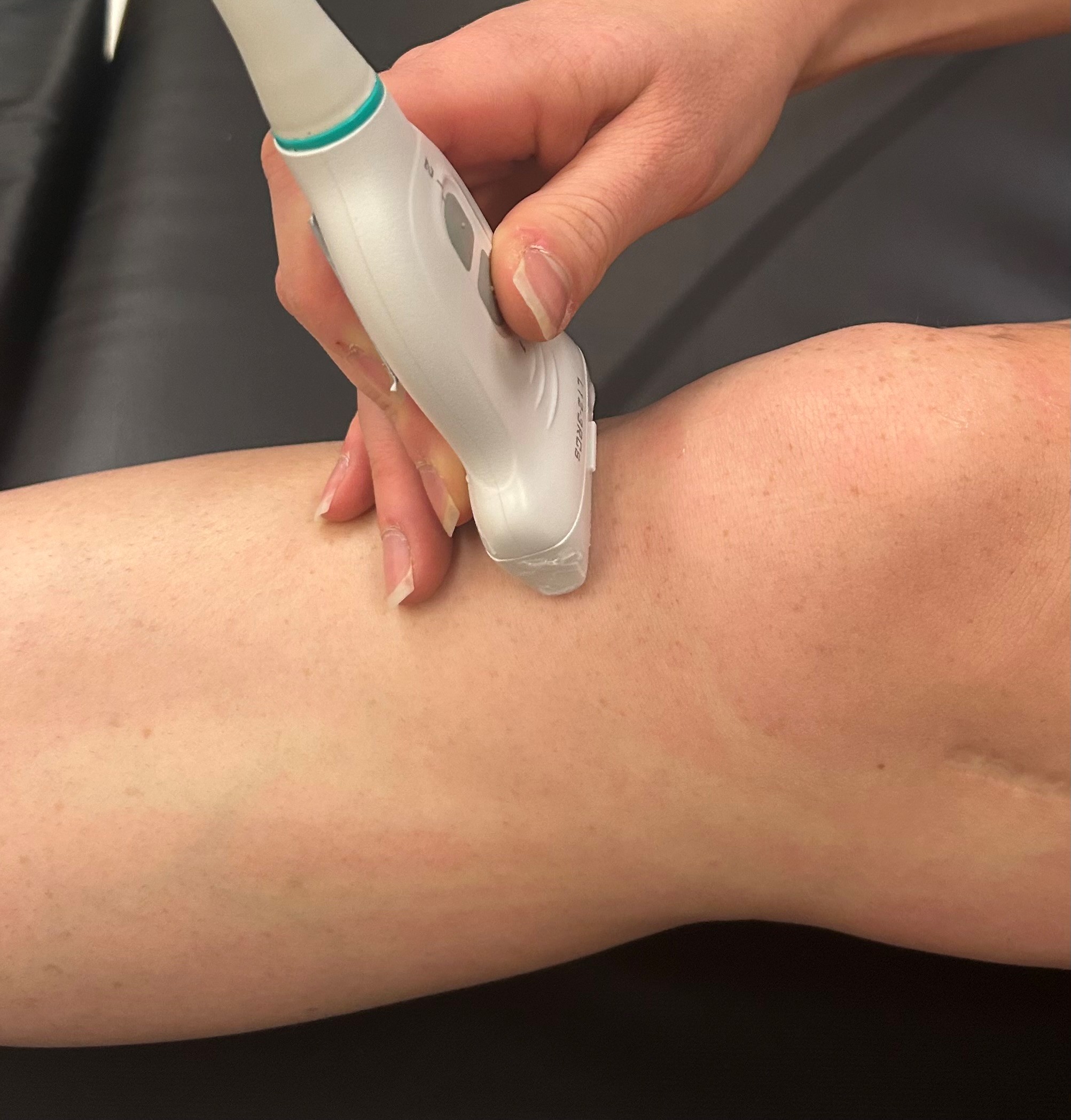
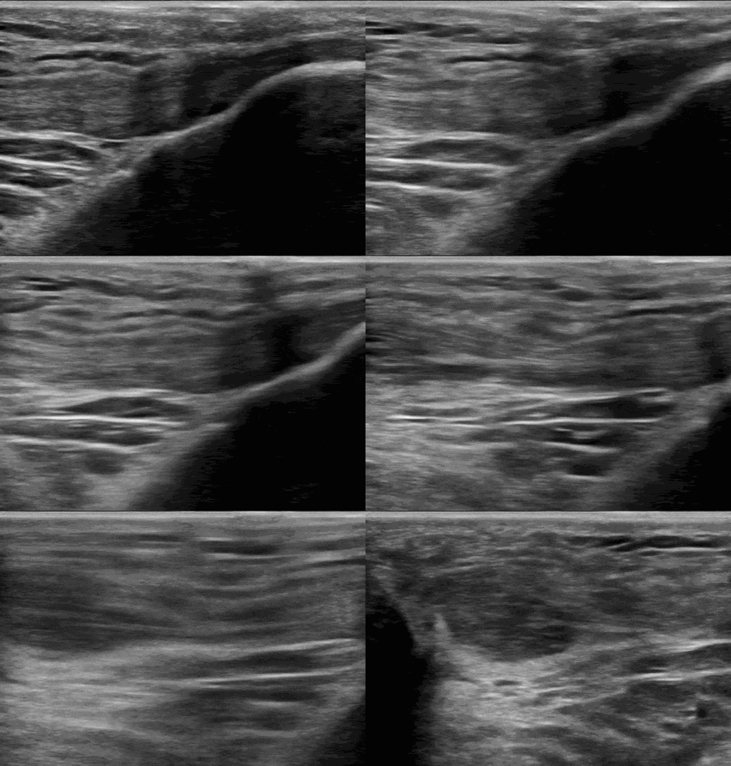
Have the patient lay supine with the knee exposed. Use the linear transducer in sagittal orientation with the probe marker facing cranially. Scan from the patella to visualize insertion, to the tibia. Switch to transverse orientation with the probe marker facing the patient’s right and repeat. Scan from insertion at the patella to the tibia.
Pathologic Patellar Tendon
Sagittal scanning
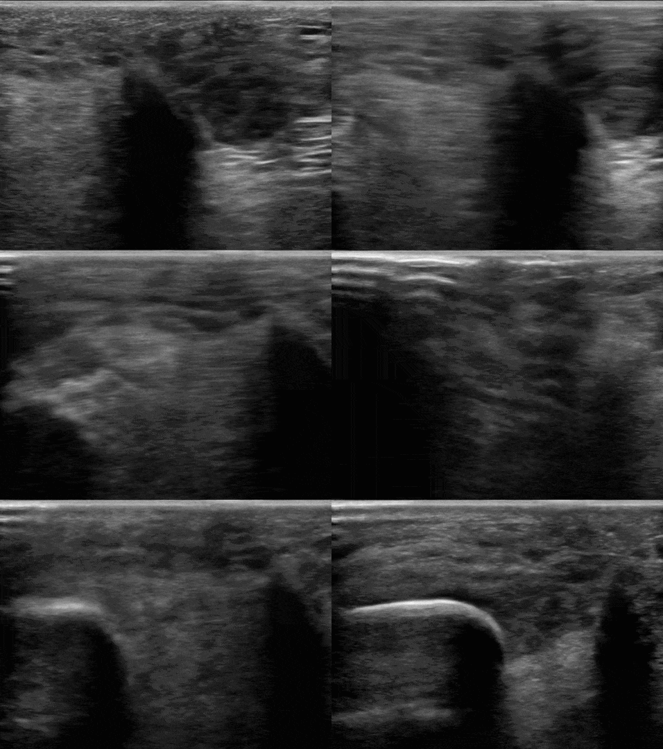
Transverse Scanning
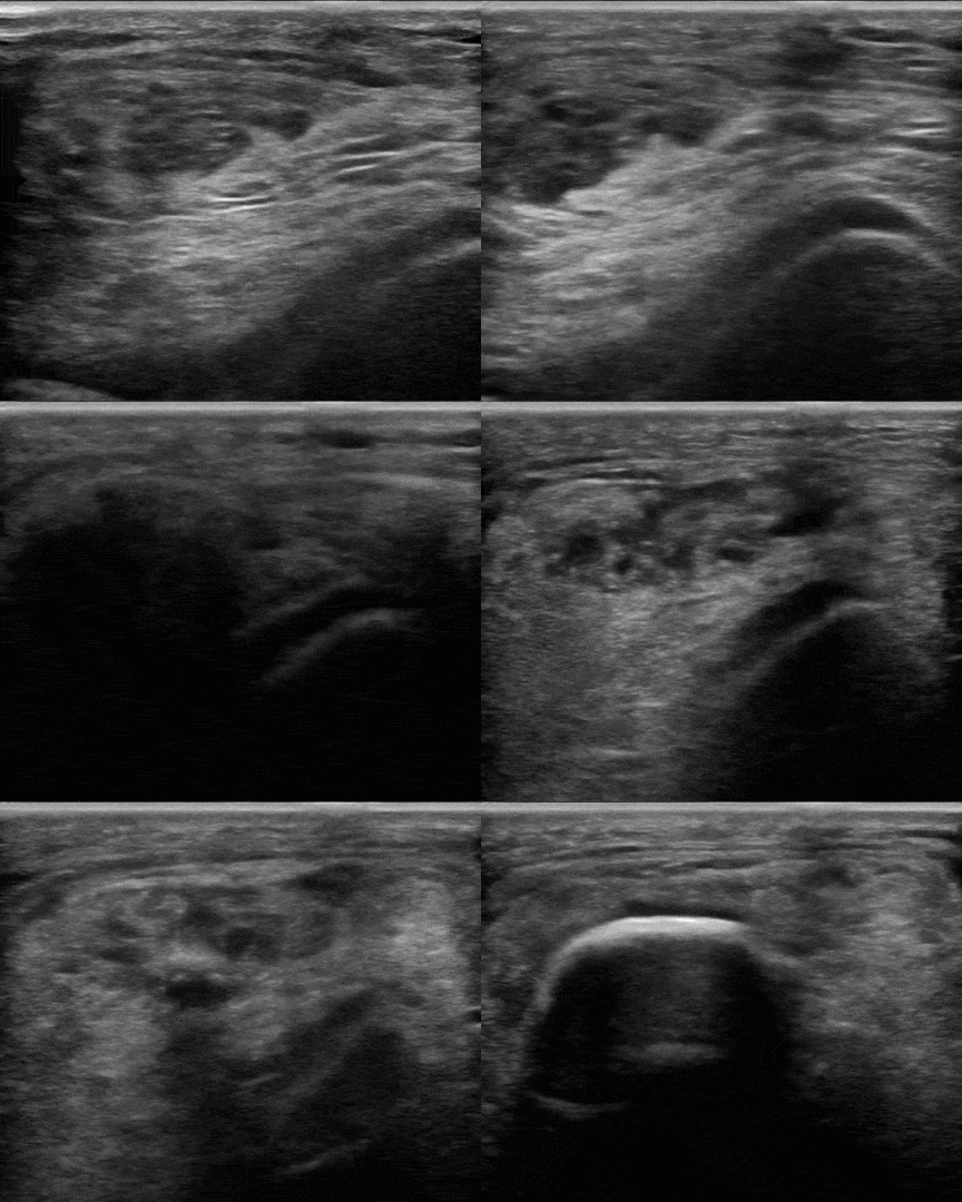
Try to identify the structures demonstrated in the below images.
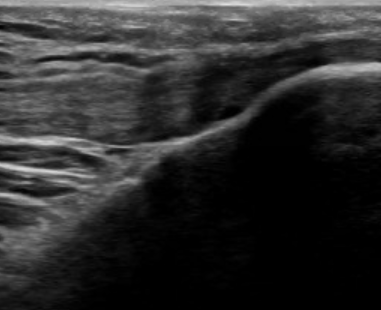
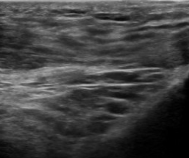
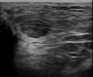
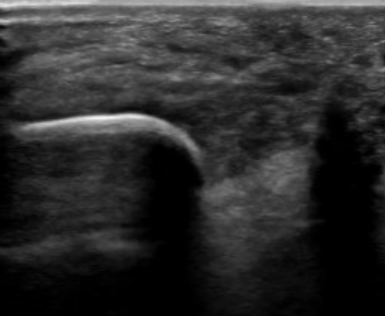
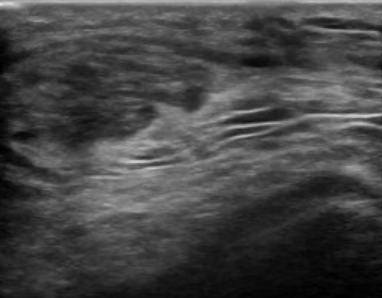
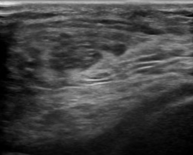
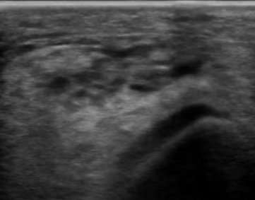
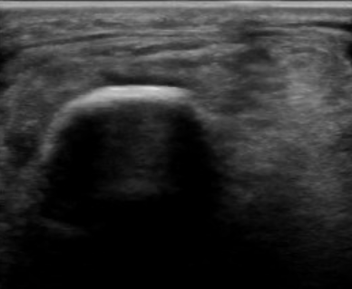
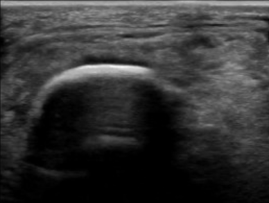
Click here to see labeled images.
Physical Exam/Special tests:
The patient will have a palpable defect or tenderness inferior to the patella. The patella may be “high riding” or sit more proximally than expected over the distal femur. An effusion may be present. The patient will have painful or incomplete hip flexion and knee extension.
Visit our case file for more information about the presentation and management of patellar tendon rupture!
PATELLAR TENDON CASE FILE
Written by: Meghan Dillan, MD
Special thanks to Dr. David Haidar for the provided ultrasound images.
References:
- Brinkman JC, Reeson E, Chhabra A. Acute Patellar Tendon Ruptures: An Update on Management. J Am Acad Orthop Surg Glob Res Rev. 2024;8(4):e24.00060. Published 2024 Apr 3. doi:10.5435/JAAOSGlobal-D-24-00060
- Mabrouk A, Siwiec RM. Patellar Tendon Rupture. In: StatPearls. Treasure Island (FL): StatPearls Publishing; February 15, 2026.
- Steele R, Hayden SR, Ward N, Macias M. Patellar Tendon Rupture Bedside Diagnosis. The Journal of Emergency Medicine. 2021. doi:10.1016/j.jemermed.2020.09.051