Shoulder Case 1 Diagnosis
Posterior Shoulder Dislocation
Diagnosis
Adequate radiographs are essential for diagnosis and associated fractures, which are common. Posterior shoulder dislocations may appear deceptively normal on AP or Grashey views.
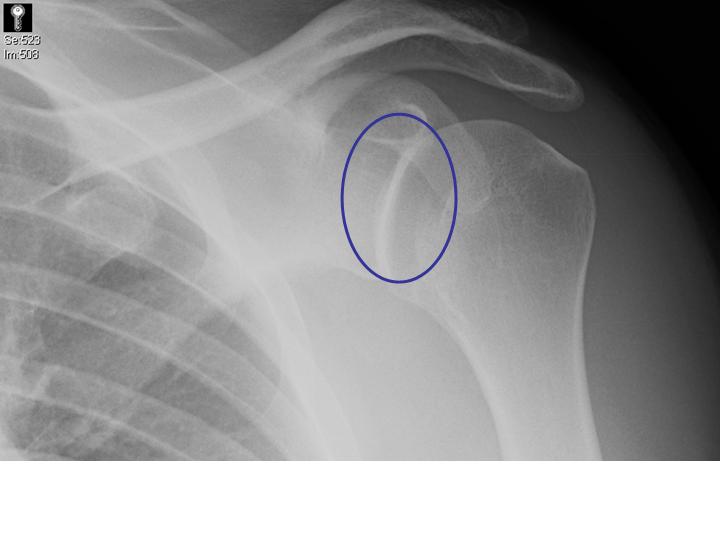
- The normal elliptically-shaped overlap of the humeral head and the glenoid may be absent in posterior shoulder dislocations.
- The articulation may be mistaken as normal, but part of the glenoid may actually be vacant, the so called "rim sign."
- In some cases, the humeral head is rotated in such a manner that the beam goes through both the greater and lesser tuberosity, attenuating the humeral head, giving it a "hollowed-out" or "cystic" appearance (see additional images).
The patient's AP film (left) shows an abnormal glenohumeral articulation (no elliptical glenohumeral overlap)without a "rim sign" or "cystic" humeral head.
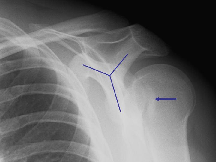
Despite the subtleties of interpreting the AP radiographs, the diagnosis should be apparent with adequate scapular "Y" or axillary views, so at least one of these must be routinely ordered. Notice how the humeral head lies posterior to the glenoid in the scapular "Y" view, making the diagnosis easier. The arrow points to the impaction fracture of the humeral head from impaction against the glenoid.