Pelvis/Hip Case 1 Diagnosis
Avascular Necrosis of the Femoral Head
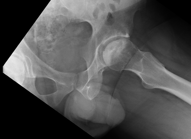
Plain radiographs may be unremarkable in the early stages of AVN (stage I) or show osteopenia.
In mild disease (stage II), plain films may show a mixed picture of osteopenia and sclerosis and may also have subchondral cysts. The femoral head maintains its normal contour (as in the patient in this case).
In moderate AVN (stage III), sclerosis and a linear subchondral lucency referred to as a "crescent sign" may be visible. There may be irregularity of the femoral head from cortical collapse.
In more advanced disease (stage IV), there is collapse of the femoral head and joint space narrowing or destruction from secondary arthritis.
MRI is more sensitive (>90%) and is able to define the deformity of the articular surface as well as subchondral edema.