Bone Lesions Case 2 Diagnosis
Benign Bone Lesions
Diagnosis
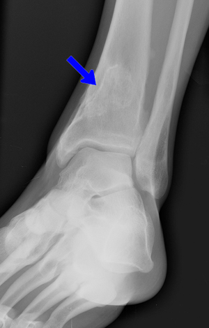
NOF is diagnosed on the basis of characteristic appearance on standard radiographs. NOFs occur as solitary lesions at the metaphysis of long bones (typically distal femur or proximal/distal tibia) with a characteristic multiloculated or 'bubbly'appearance with surrounding sclerosis.
Osteochondromas(the 2nd most common benign bone tumor) present as palpable masses at the metaphysis of long bones, most typically around the knee. Radiographically they appear as compact, pedunculated, cartilage-capped growths.
Osteoid osteomas are small, solitary lesions that are often intensely painful; they are poorly visualized on plain films and often require CT for diagnosis. The characteristic appearance is that of a 'target sign' with a radiolucent nidus surrounded by a sclerotic rim.
Bone cysts are usually found in the proximal femur or humerus and appear as simple, fluid-filled lesions of the metaphysis/diaphysis and produce no palpable deformity.
Radiographic features that suggest a malignant lesion include:
- poorly defined margins
- gross deformity of bone
- multiple lesions
- evidence of bony destruction (especially in combination with rapid new-bone formation).