Pelvis/Hip Case 2 Diagnosis
Pelvic Fractures
Diagnosis
Standard radiographic examination of the pelvis includes an AP view, followed by CT depiction of documented fractures.
Supplemental views which may be requested by consultants include:
- inlet view (beam directed 60 degrees toward the feet) to assess posterior arch
- outlet view (beam directed at 30 decrees toward the head) to look at anterior arch.
These supplemental views are seldom needed when CT has been performed.
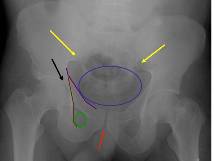
When interpreting plain films, look at the:
- illiopubic lines (purple line)
- illioischial lines (brown line)
- acetabulum (black arrow)
- pelvic inlet (blue oval)
- obturator foramina (green circle)
- sacroiliac joints (yellow arrows)
- pubic symphysis (red arrow).
Make sure all arcs and rings and smooth and continuous without stepoffs. If unsure, compare to the other side for symmetry. If the ring is not intact in one spot, examine carefully for the second break in the ring. Clues to posterior arch disruption include L5 transverse process fracture, ischial spine or lower sacral avulsion, and asymmetry of sacral fora
mina.
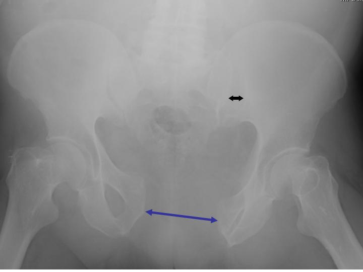
Widening of SI joints > 4mm is abnormal (black arrow). Symphyseal widening > 5mm is abnormal (blue arrow).