Foot Case 4 Additional Images
Calcaneus fracture with loss of Bohler's angle
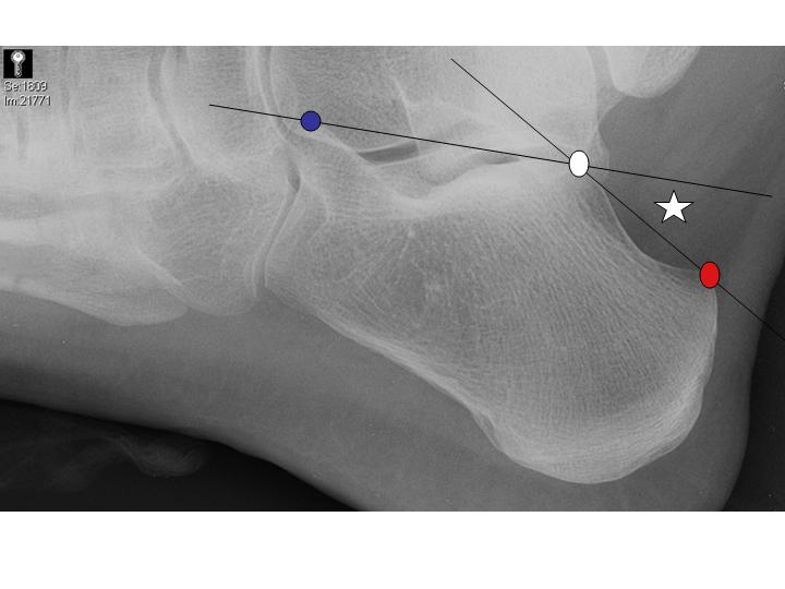
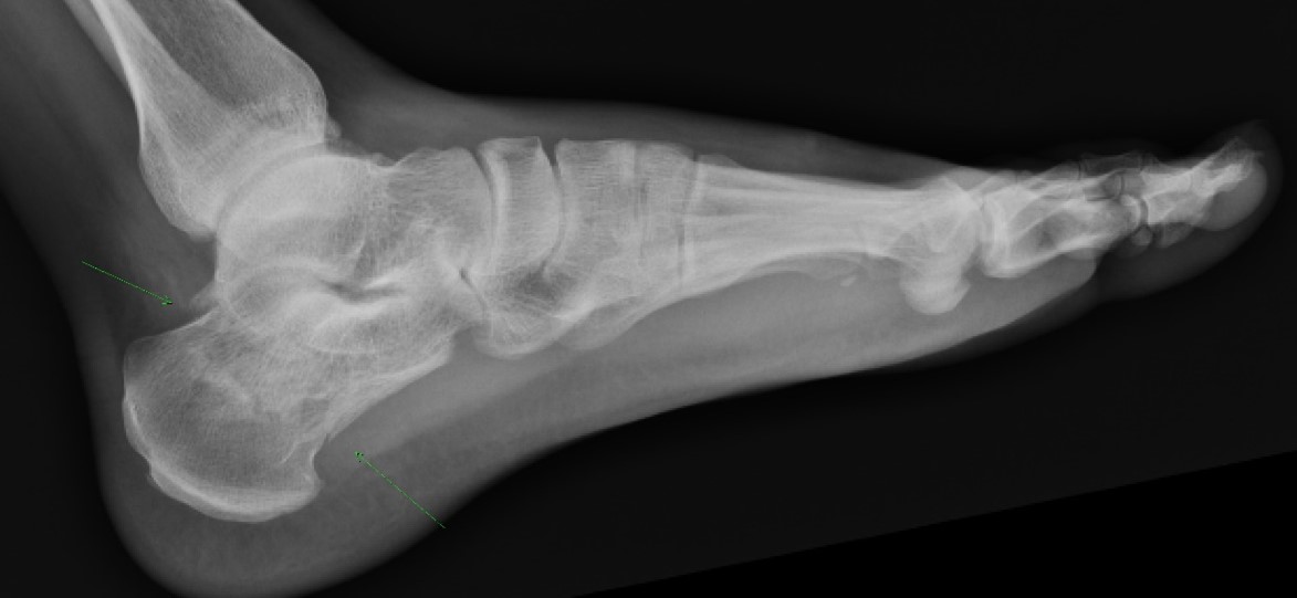
Normal Bohler's angle (star) and trabeculations in a patient with no calcaneus injury. Bohler's angle is formed by intersecting two lines. One is drawn from the most cephalic portion of the tuberosity (red dot) to the highest point of the posterior facet (white dot). The other line is drawn from the highest point of the posterior facet (white dot) to the most cephalic part of the anterior process (blue dot).
The arrow points to the fracture of the peroneal trochlea of the calcaneus in the case presented above.
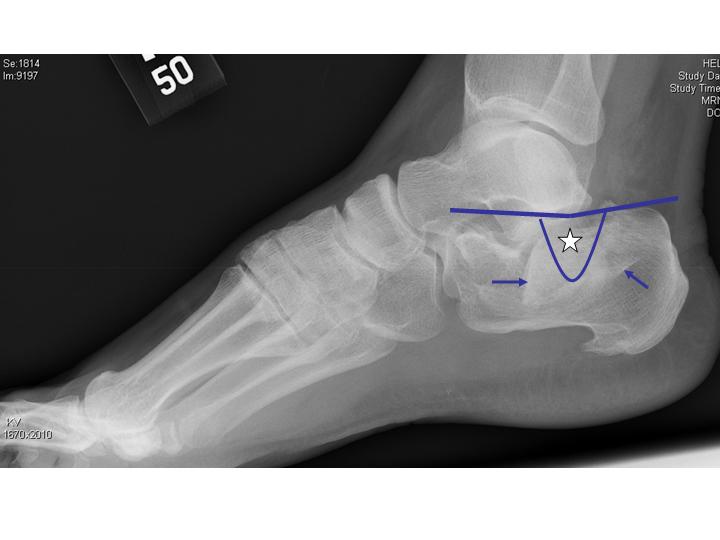
Same patient, x ray magnified. Note the loss of Bohler's angle (star), which is actually negative. There are multiple linear areas of sclerosis that cross trabecular lines (arrows).
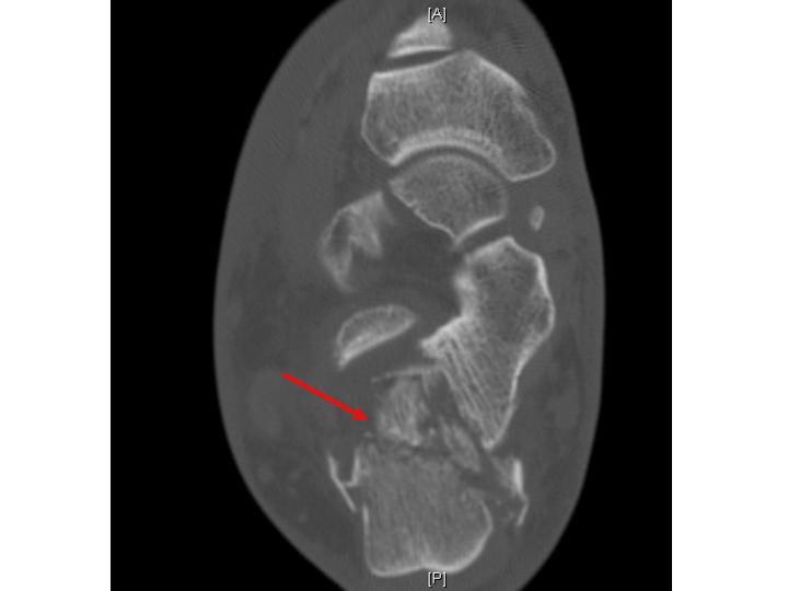
CT of the patient showing the severely comminuted calcaneal fracture. Notice how much more appreciable the comminution is on CT compared to plain films.
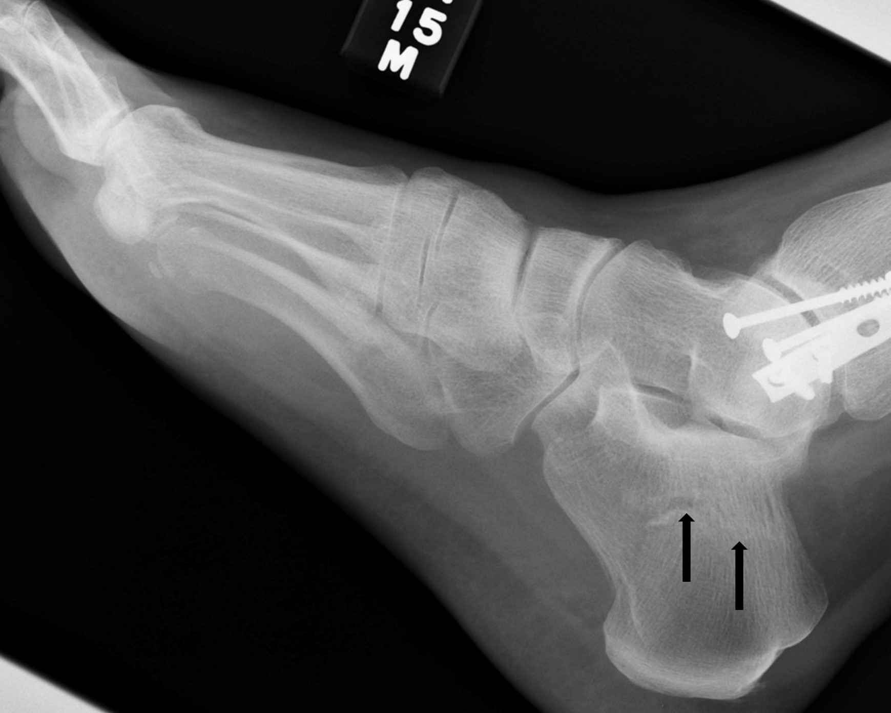
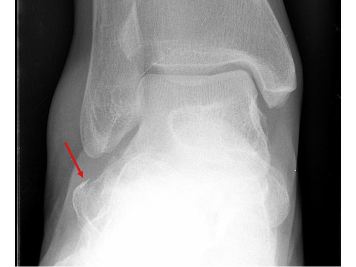
This image shows a different patient who has a subtle calcaneus fracture that is revealed by the sclerotic disruption of the normally smooth continuous trabeculae.
The above image displays a calcaneus fracture with more obvious disruption of the smooth trabecular pattern.
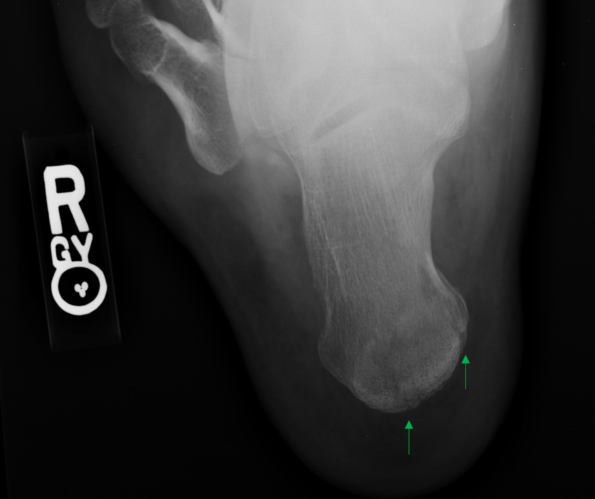
The above patient's calcaneus fractures are evident on this “calcaneal view”, with the beam aimed axially up through the heel.